Maulana Azad National Institute of Technology (MANIT) is one of the leading institutions of national importance in the area of technical education, established with the objective of developing a "Centre of Excellence" in the central region. It aims at becoming a multi-disciplinary Centre for technical education by strengthening both teaching and research activities besides contributing to the needs of rural community, society and industry at large. Institute is fully funded by Ministry of Human Resource Development, Government of India and is governed as per provisions made under the National Institute of Technology, Science Education and Research Act 2007 (NITSER Act 2007).
MANIT is on the way to develop Central Research Facility aiming to create advanced research facility with more emphasis on Industry-academia interaction and to meet community needs for technology driven solutions. These state-of-the-art analytical and high-end instruments are housed at a single location manned by qualified & skilled personnel to provide scientific and technical services for supporting research carried out by UG, PG, PhD and project students and also for consultancy works. Such a facility represents a key commitment for preserving and raising the efficiency of research to top international standards. It strengthens the research-relevant infrastructure of basic science and technologyis located in New Teaching Block (NTB) It house the most modern equipments as a facility to the users of the ever-expanding number of UG, PG students & researchers with various needs coming from different fields.
- To create state of the art research facilities for promotion of interdisciplinary research in areas of Chemical Engineering, Materials Science and technology and Physics
- To make the facilities accessible to all internal, external and industry users for research and development.
- To promote active collaboration with institutes, universities, research laboratories and industries.
- To provide a platform for carrying out sophisticated experiments in the area of materials science and engineering.
The Central Research Facility envisions are to provide state of the art materials characterization systems for all students at MANIT Bhopal and other academic and industry users to carry out their research activities. It further envisions the creation and maintenance of appropriate ambiance entailing drives in research for the development of appropriate materials technology. The equipments housed in the CRF are used for various types of characterization including study of structure and chemical composition of materials at different length scales (sub-nanometre to millimetre), phase transitions, as well as evaluation of mechanical, electrical and optical properties.

Dr. Savita Nema
Coordinator, CRF & Professor
Department of Electrical Engineering
More Information

Dr. S. Suresh
Convenor & Faculty In-charge, CRF
Associate Professor, Department of Chemical Engineering
More Information
-
Dr. Rajesh Purohit, Professor, Mechanical Engineering Department
-
Dr. Fozia Z Haque, Associate Professor, Physics Department
-
Dr. Raman Nateriya, Assistant Professor, Mechanical Engineering Department
-
Dr. Vijay Kumar Bulasara, Associate Professor, Chemical Engineering Department
-
Dr. Amit Ojha, Associate Professor, Electrical Engineering Department
-
Dr. C. Sasi Kumar, Associate Professor, Metallurgical and Material Engineering Department
-
Dr. Khushhali M. Pandey, Associate Professor, Biological Science and Engineering
-
Dr. Bharat K. Modhera, Associate Professor, Chemical Engineering Department
-
Dr. Dharmendra Pandey, Assistant Professor, Chemical Engineering Department
-
Dr. Dharmendar Sharma, Assistant Professor, Chemistry Department
-
Dr. Piyush Kumar Patel, Assistant Professor, Physics Department
-
Dr. Jyoti Rani, Assistant Professor, Physics Department
-
Dr. Rupak Kishore, Assistant Professor, Chemical Engineering Department
-
Dr. Ram Kishore, Assistant Professor, Metallurgical and Material Engineering Department
-
Dr. Sunder Lal Pal, Associate Professor, Chemical Engineering Department
-
Dr. Sanjay Srivastava, Professor, Metallurgical and Material Engineering Department
-
Dr. Sukanti Behera, Assistant Professor, Chemistry Department
-
Dr. Arvind Kumar, Assistant Professor, Mechanical Engineering Department
-
Dr. Narendra Laxman Gajbhiye, Assistant Professor, Mechanical Engineering Department
-
Dr. Sudhanshu Kumar, Assistant Professor, Mechanical Engineering Department
-
Dr. Rutuja M. Chavan, Assistant Professor, Civil Engineering Department

Mr. Prashast Manglik
Technical Officer
Email id:- prashast[AT]manit.ac.in
Phone No:-0755-405-1006
-
Mr. Harish Vaidya, Deputy Registrar (Store & Purchase)
-
Mr. Rakesh Singh (Storekeeper)
Electrochemical Impedance Spectroscopy (EIS) with Solar Simulator
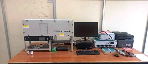
Electrochemical Impedance Spectroscopy (EIS) with Solar Simulator is an advanced characterization technique used for studying the electrochemical and photovoltaic properties of various materials, thin films, and devices. This setup is particularly useful in the field of solar cell research, corrosion science, batteries, fuel cells, and super capacitors.
The integrated Solar Simulator provides a controlled and calibrated light source (AM 1.5 G spectrums), allowing researchers to simulate real sunlight conditions while performing EIS measurements. This enables accurate evaluation of device performance under standard testing conditions.
Instrument Details:- EIS – Auto Lab
Solar simulator – ScienceTech
Key Features:-
- Wide frequency range impedance analysis.
- High-precision potentiostat/galvanostat integrated with solar simulator.
- Illumination with AM 1.5G spectrum, calibrated intensity (100 mW/cm²).
- Suitable for solid, liquid, thin film, and device-level studies.
Applications:-
- Photovoltaic devices (solar cells – perovskite, organic, dye-sensitized, silicon-based).
- Electrochemical energy storage systems (Li-ion batteries, super capacitors).
- Corrosion studies and protective coatings.
- Photo electrochemical water splitting and hydrogen production.
- Semiconductor/electrode interface characterization.
Characterization charges of EIS with solar simulator at CRF
|
EIS with solar simulator |
|||||
|---|---|---|---|---|---|
|
Name of the equipment/ Testing |
MANIT Users |
Other Educational Institutes (Rs.) |
Govt. R&D Institution /Labs (Rs.) |
Industry
(Rs.) |
|
|
Non-funded (Rs.) |
Funded (Rs.) |
||||
|
EIS with solar simulator |
Rs. 100/- Per Sample
|
Rs. 200/- Per Sample |
Rs. 500 /- Per Sample |
Rs. 1000/- Per Sample |
Rs. 2000/- Per Sample |
Confocal Raman Microscope

Raman spectroscopy is an analytical technique where scattered light is used to measure the vibrational energy modes of a sample. It is named after the Indian physicist Dr. C.V. Raman who, together with his research partner K.S Krishnan, was the first to observe Raman scattering in 1928. Raman spectroscopy can provide both chemical and structural information, as well as the identification of substances through their characteristic Raman ‘fingerprint’. Raman spectroscopy extracts this information through the detection of Raman scattering from the sample.
Confocal Raman Microscopy: Confocal Raman Microscopy combines the spectral information from Raman spectroscopy with the spatial filtering of a confocal optical microscope for high resolution chemical imaging of samples. The spectral Raman information is sensitive to the vibrational modes of the sample and provides extensive chemical, physical and structural insight, while the confocal optics of the microscope enables the analysis volume within the sample to be spatially filtered with high resolution in both the lateral (XY) and axial (Z) axes. The synergy between the spectral information enables the chemical analysis of individual particles, discrete sample features or layers down to less than 1um in size.
Model Name – CTR 300-S/C-x
Make: TechnoS Instruments, Jaipur India.
The Confocal Raman Microscope consists of two lasers i.e. 532nm and 785nm for Raman characterization. Other items to the above are aberration corrected Czerny-Turner single spectrograph, 3 gratings for CTR 300. Window based computer with a data collection and processing software. Entrance slit: 10 μm - 3.0 mm, RS232C/USB, optical fibre of 2m. Spectra repeatability <0.1cm-1, Spectra Scan linearity <0.5cm-1. Spectrum Range: 200 – 2100 nm, Raman Shift: 50 – 5000cm-1.
Optical Microscope ST-/LV/Ti (INV)
Confocal Raman optical microscope with < l/2 spatial resolution (<500nm standard using x100) objective lens, Raman probe with Raman Filter set, halogen light (ref/trans), Objective lens : x5, x10, x20, x50, x100 (Oil immersed) & CCD colour video camera variable spot size up to 300 μm (WAC).
Imaging Spectrograph
Focal Length Spectral Resolution Gratings Raman Range
CTR 300-S/C-x F/4.1 300 <0.4 cm-1/pixel 3 50-5000 cm-1
Characterization charges of Confocal Raman Microscope at CRF
|
Confocal Raman Spectrometer |
|||||
|---|---|---|---|---|---|
|
Testing Name |
MANIT Users |
Other Educational Institutes (Rs.) |
Govt. R&D Institution /Labs (Rs.) |
Industry (Rs.) |
|
|
Non-funded (Rs.) |
Funded (Rs.) |
||||
|
Confocal Raman Spectrometer(only spectra) |
Rs. 100/- Per Sample
|
Rs. 200/- Per Sample |
Rs. 400/- Per Sample |
Rs. 800/- Per Sample |
Rs. 1000/- Per Sample |
|
Confocal Raman Spectrometer (Spectra, PL & Mapping) |
Rs. 300/- Per Sample
|
Rs. 600/- Per Sample
|
Rs. 2000/- Per Sample |
Rs. 5000/- Per Sample |
Rs. 8000/- Per Sample |
Field Emission Electron Microscope

Field Emission Scanning Electron Microscopy (FESEM) is a technique that uses electrons to create images of materials and study their surface topography. FESEM is also known as a cold cathode field emitter because it doesn't use heat.
Basic principle:
The technique in which electrons are used in place of light to take the image is known as electron microscopy. Electron microscopy is of two types, i.e., scanning electron microscopy (SEM) and field emission scanning electron microscopy. FESEM differs from SEM in the type of emitter; in SEM, a thermionic emitter is used, while in FESEM, a field emitter is used.
As the name suggests, thermionic emitters use heat to eject the electrons, and when the heat overcomes the work function, electrons are ejected, but the intensity is low and materials evaporate easily. In the field emitter ejection, these limitations are removed. In FESEM, the filament is placed in the high electric potential gradient to generate the electrons.
Generally, tungsten (W) wire is used for the production of electrons. This technique does not use heat so it is also known as a cold cathode field emitter. The electron beam is focused on the sample and the beam obtained after electron-sample interactions contains all the information related to chemical composition, morphology (texture), crystalline structure, and orientation of the material. It is one of the best techniques to uncover the surface structure, morphology, and composition of the sample. It provides a high-quality three-dimensional surface image of the material with high resolutions. The advantage of high resolution is that the closely lying different particles can be easily pointed out by enhancing the magnification.
Energy dispersive spectroscopy
Energy-dispersive X-ray spectroscopy (also known as EDS, EDX, or EDXA) is a powerful technique that enables the user to analyze the elemental composition of a desired sample. The major operating principle that allows EDS to function is the capacity of high energy electromagnetic radiation (X-rays) to eject 'core' electrons (electrons that are not in the outermost shell) from an atom. This principle is known as Moseley's Law, which determined that there was a direct correlation between the frequency of light released and the atomic number of the atom.
Removing these electrons from the system will leave behind a hole that a higher energy electron can fill in, and it will release energy as it relaxes. The energy released during this relaxation process is unique to each element on the periodic table, and as such bombarding a sample with X-rays can be used to identify what elements are present, as well as what proportion they are present in.
Shown below is an example of how EDS works. The letters K, L, and M refer to the n value that electrons in that shell have (K electrons, closest to the nucleus, are n=1 electrons), while α and β indicate the size of the transition. The relaxation from M to L or L to K are therefore described as Lα or Kα, while going from M to K would be a Kβ transition. The means that are used for describing these processes as a whole are known as Siegbahn notation.

● Energy dispersive spectroscopy (EDS) detector is attached to the system to find out the composition of the samples which uses eZAF correction.
● Element detection range of EDS varies from Be (4) - Am (253) with 70mm2 SDD window with peltier cooling.
Instrument details
Instrument Model: CLARA
Make of FESEM: TESCAN, Czech Republic
Specifications:
- Accelerating Voltage: 0.2keV - 30keV
- Lens Configuration: Condenser Lens – 2 Stage, IML – 1 stage, Objective Lens – 1 Stage.
- Magnification: 20X – 2000KX
- SEI Resolution: 0.9nm @ 15KeV
Sample preparation for FESEM:
·Medium for dispersion Ethanol /Methanol / Water /Iso-propyl alcohol. Any other medium should be provided by the user. Dispersion will be done by ultrasonication.
· Kindly mark the edge of the sample to be observed for Cross section.
·Base of the sample should be flat for mounting on sample holder.
·Biological samples will be accepted only after primarily fixation with suitable fixative.
·Samples should be in dry form. Hydrated samples must be dried properly before sending.
·Sample preparation if any should be done at user end (cutting the sample for CS, freeze fracturing, sample fixation for biological samples, staining of samples, oven drying should be done by the user).
·The samples should withstand high vacuum (~ 10 -5 Pa). Wet samples can’t be done in FEG-SEM
·Any solid structures like in FESEM within a Maximum range of sample size of approx 12.5mm dia and 80mm height is acceptable. Bring Fresh CD for FESEM which should not be used for other equipment’s data.
Characterization charges of Field Emission Electron Microscope (FESEM) at CRF
|
Field Emission Electron Microscope(FESEM) |
|||||
|---|---|---|---|---|---|
|
Name of the equipment/ Testing Mode |
MANIT Users |
Other Educational Institutes (Rs.) |
Govt. R&D Institution /Labs (Rs.) |
Industry
(Rs.) |
|
|
Non-funded (Rs.) |
Funded (Rs.) |
||||
|
FESEM |
Rs. 200/- Per Sample
|
Rs. 400/- Per Sample |
Rs. 1200 /- Per Sample |
Rs. 2000/- Per Sample |
Rs. 3000/- Per Sample |
BET Surface Area Analyzer

Instrument model: BELSORP- MINI X (BELSORP – series) from Microtrac BEL Corp
Brunauer-Emmett-Teller (BET) surface area analyzer is a non-destructive technique that is based on the BET theory which demonstrates the physical adsorption of the gas molecules (adsorptive) on a solid/porous surface (sample). BET theory is an extension of the Langmuir theory from monolayer adsorption to multilayer adsorption of adsorptive onto the adsorbent. In this technique, an inert gas such as nitrogen flows continuously over a solid sample which is suspended in a defined gaseous volume through the sample cell. Now, the small molecules of the inert gas adsorb to the surface of the sample and in its inner pore’s surface through Vander Waals forces. The amount of gas adsorbed depends mainly on the exposed surface area of the materials and also on the factors such as temperature, gas pressure, and degree of strength of interaction between the gas molecules and solid surface. The rate of adsorption can be used to calculate the specific surface area of a solid material. This instrument is capable to measure the adsorption/desorption isotherms, surface area (Langmuir and BET), pore size (BJH method), pore size distribution, and pore volume of the sample.
BET specific surface areas from 0.01 m2/g ~ (N2) and pore size distribution from 0.7 ~ 500 nm (option: 0.35 ~ 500 nm by molecular probe method). Up to 3 specimens can be measured simultaneously, enabling the measurement times for multiple sampl
Instrument model: BELCAT II from Microtrac BEL Corp.
The chemisorption process involves the creation of strong electronic bonds between adsorbate molecules and certain surface locations, which are known as surface active sites. Chemisorption is irreversible and involves more heat than physical adsorption. Chemisorption catalyst analyser BELCAT can evaluate catalysis by using the following methods; Temperature Programmed Desorption spectrum measurement (TPD), Temperature Programmed Reaction spectrum measurement (TP Reaction), Temperature Programmed Reduction spectrum measurement (TP Reduction), Temperature Programmed Oxidation spectrum measurement (TPO), Metal dispersion measurement, Pulse injection measurement, and Single-point BET method to measure the specific area of samples. The BELCAT can be programmed automatically to do pre-treatment of the samples, measurement, and calculations. The study related to the acidity and basicity of the solid samples can also perform with this instrument.
Sample specifications
1. Only solid porous samples (powder) are allowed in the equipment.
2. 100 mg (minimum)
(Suggestion: please carry enough sample)
Limitation
1. Liquids samples are not allowed to run.
2. Samples should not melt or deteriorate inside the cell holder during characterization.
Characterization charges of BET Surface Area Analyzer at CRF
|
BET Surface Area Analyzer |
|||||
|---|---|---|---|---|---|
|
Name of the equipment/ Testing Mode |
MANIT Users |
Other Educational Institutes (Rs.) |
Govt. R&D Institution /Labs (Rs.) |
Industry
(Rs.) |
|
|
Non-funded (Rs.) |
Funded (Rs.) |
||||
|
Brunauer - Emmett - Teller (BET) |
Rs. 200/- Per Sample
|
Rs. 400/- Per Sample |
Rs. 1000/- Per Sample |
Rs. 2000/- Per Sample |
Rs. 4000/- Per Sample |
es to be shortened significantly & up-to 6 samples can be degassed simultaneously at temperature from Rt to 400 Deg C.
Gas Chromatography/ Mass Spectrometry (GCMS/MS)

Gas chromatography – mass spectrometry (GC-MS/MS) is an analytical method that combines the feature of gas – chromatography and mass spectrometry to identify different substances within a test sample. Applications of GC-MS include drug detection, fire investigation, environmental analysis, explosives investigations, food and flavor analysis, and identification of unknown samples, including that of material samples obtained from planet Mars during probe missions as early as the 1970s. GC – MS can also be used in the airport security to detect substances in luggage or on human beings. Additionally, it can identify trace elements in materials that were previously thought to have disintegrated beyond identification. Like liquid chromatography – mass spectrometry, it allows analysis and detection even of tiny amounts of a substance.
Instrument Model: GCMS-TQ8050 NX, Make: Shimadzu
Sample preparation:
For GC-MS sample preparation, user has to use a simple filtration technique to remove solid particles and inorganic salts if you are using a reaction mixture. The solution must be very diluted (0.1 mg/mL). It is recommended that your analyte must have a molecular weight of less than 500 g/mol. Only organic compounds are allowed for characterization in GCMS.
|
INSTRUCTIONS FOR GCMS USERS |
|
1. Only volatile and thermally stable compounds can be analyzed. 2. Samples can be injected in liquid form only, if solid sample is not soluble in solvent then the sample cannot be injected. Samples in diluted form can be injected, if high in concentration, please make it dilute. 3. The sample should be dissolved in volatile organic solvents, preferably hexane or isooctane. Other solvents like, methanol, ethanol, ethyl acetate etc. can also be used. All volatile solvents are acceptable except such as Glycols, PEG etc. You can use DMSO & THF as diluents. The sample should be free of particulates of size > 0.45 μ 4. One sample refers to one injection. Multiple injections of the same sample will be treated as multiple samples & samples above C40 can only be used after extraction only. 5. Un-dissolved Samples with particles, Hazy samples, water sample & thick liquid samples cannot be injected in the equipment. 6. Before and after Run sequence of samples, always do column conditioning, during conditioning keep the filament OFF. |
Characterization charges of GCMS/MS at CRF
|
GCMS/MS |
|||||
|---|---|---|---|---|---|
|
Name of the equipment/ Testing Mode |
MANIT Users |
Other Educational Institutes (Rs.) |
Govt. R&D Institution /Labs (Rs.) |
Industry
(Rs.) |
|
|
Non-funded (Rs.) |
Funded (Rs.) |
||||
|
GCMS/MS |
Rs. 100/- Per Sample
|
Rs. 200/- Per Sample |
Rs. 1000/- Per Sample |
Rs. 1800/- Per Sample |
Rs. 2000/- Per Sample |
Note: In addition to the above charges, GST @ 18% will be charged extra.
Contact Person: Mr. Prashast Manglik
Technical Officer, CRF
Email Id: prashast@manit.ac.in
Office Tel: +91- 755-4051006
(Goods and Service Tax 18 % w.e.f. 1st July 2017)
No Postal Charges for any analysis.
- Dispatch address will be same as address on letter head.
- GST is applicable to all the users and is calculated as per current rate 18% (w.e.f. from 1st July 2017).
- All the samples should accompany the payment receipt, in order to be considered for booking. All the payments should be in favour of "ICSC, MANIT Bhopal". In case of electronic transfer details must be submitted to the account section for verification.
- The payment should be rounded off the nearest figure (integer value).
Example: If total charges (including tax etc) comes out to be Rs.169.67/- (i.e. greater than or equal to 50 paisa) then the amount should be rounded off to Rs.170/- but if the total charges (including tax etc) comes out to be Rs.169.47/-(i.e. less than 50 paisa) then the amount should be rounded off to Rs.169/- - Requesting letter exactly as it is: "Content of this report/bill is meant for our information only and we will not use the content of this report for advertisement, evidence, litigation or quote as certificate to third party". The technical report is conforming to the sample provided by the party, and this report is not submitted for any court of law.
- Results will be available either in softcopy sent through e-mail or hardcopy needs to be collected from CRF office. Results will not be sent through post.
- All the users are required to submit MSDS (Material Safety Data Sheets) for each sample they submit and a certificate stating that the samples submitted are non- toxic / non-hazardous inflammable and that the sample does not require special precaution while handling. Email sent to Coordinator/Members/Others will not be entertained.
Booking System-The Process
- Please click here for confirmation of charges before making payment.
- Any wrong information quoted may lead to cancellation of your booking.
- Please submit payment receipt, samples, equipment requisition form & the covering letter on organisation’s letter head.
- The Equipment Requisition form for samples analysis duly signed by the concerned Supervisor & HoD along with seal and signature, with details of NEFT/RTGS/UPI transfer and GST number of the concerned Institute, may kindly be sent at the address given below along with the e-mail and mobile number in the application. The equipment requisition form can be downloaded from the website of the institute.
- Kindly do not dispatch the samples without prior discussion with the facility in-charge and contact the facility in-charge before sending the samples for measurement.
- Mode of the payment:
The payment should be made in advance as under: The mode of the payment is strictly by online transfer through NEFT/RTGS/UPI. Demand draft is no longer accepted. The details of the account are as under:
Name of the Account holder : MANIT, ICSC Bhopal
Name of the Bank : SBI, MANIT Campus Bhopal
Account No : 10020150016
IFSC Code : SBIN0001608
MICR Code : 462002014
GST Number : 23AAATD5152E1Z9
Postal Address: The Coordinator,
Central Research Facility (TA-115) NTB,
Maulana Azad National Institute of Technology - 462003
On the top of the envelope, also mention "Samples for _______characterization.”


